| TITLE |
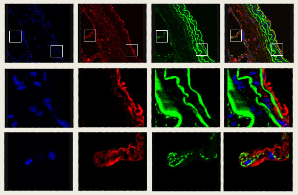
| nuclear spectrin PDE 4D4 merge |
 |
|
Fluorescent images of lung tissue taken with a NikonTi85 microscope. The second and third row of each panel are close-up sections taken from the top row (indicated in the boxes). Blue, nuclear stain. Red, fluorescent tagged antibody to non-erythroid spectrin. Green, fluorescent tagged antibody to type 4D4 phosphodiesterase. Merge, overlay of the different images. |
|
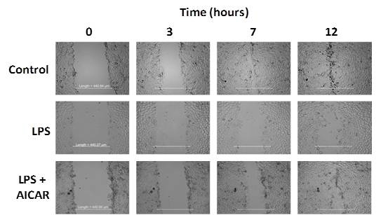
AMPK Activation Using AICAR Restores |
 |
 |
|
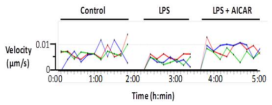
Rate of gap resealing is given in microns/sec. Velocity determined using time lapse video microscopy and Nikon Instruments moving object software. Control velocity 6.63 x 10⁻³+/- 0.46 x 10⁻³ µm/s, LPS velocity 4.44 x 10⁻³ +/- 1.22 x 10⁻³ µm/s, LPS + AICAR velocity 7.30 x 10⁻³ +/- 1.41 x 10⁻³ µm/s. |