| 2003 Case #10 |  |
|
| 2003 Case #10 Diagnosis and Discussion |
 |
|
| (Links to Other 2003 Cases are at bottom of this page) | ||
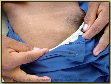
| Diagnosis: Lymphogranuloma venereum (LGV). |
 Discussion: LGV, which is due to the so-called LGV serovars (L1, L2, L3) of Chlamydia trachomatis, is a sporadic disease in North America and Europe, where it is now very uncommonly seen. In many tropical countries it is still highly endemic, including parts of Africa, India, Southeast Asia, as well as in Amazon regions of Perú and Brasil. In Iquitos, which has a large sex industry due to the simultaneous presence of military bases, oil/gas workers, and narco-traffickers, infection is highly prevalent. Diagnosis is usually made on clinical grounds as isolation in culture, PCR, LCR, antigen detection, antibody detection tests are not available. Examination of smears an biopsy material with giemsa staining for inclusion bodies is laborious and of low sensitivity. Ideally, syphilis and HIV serology as well as urethral cultures should be done, but in practice in this highly endemic and resource poor setting rarely are. Discussion: LGV, which is due to the so-called LGV serovars (L1, L2, L3) of Chlamydia trachomatis, is a sporadic disease in North America and Europe, where it is now very uncommonly seen. In many tropical countries it is still highly endemic, including parts of Africa, India, Southeast Asia, as well as in Amazon regions of Perú and Brasil. In Iquitos, which has a large sex industry due to the simultaneous presence of military bases, oil/gas workers, and narco-traffickers, infection is highly prevalent. Diagnosis is usually made on clinical grounds as isolation in culture, PCR, LCR, antigen detection, antibody detection tests are not available. Examination of smears an biopsy material with giemsa staining for inclusion bodies is laborious and of low sensitivity. Ideally, syphilis and HIV serology as well as urethral cultures should be done, but in practice in this highly endemic and resource poor setting rarely are.
Clinically, LGV presents in 3 stages. Primary LGV presents as a small painless papule (or ulcer) at the site of initial infection which may be penis, labia, vagina, or cervix. It heals spontaneously in a few days and is only noticed by the patient in 3 to 53 percent of cases. Receptive anal intercourse in males or females may result in a primary anal or rectal infection. Most often patients present in the secondary stage after the primary lesion has healed and the inguinal lymphadenopathy syndrome is the most common manifestation. However, affected nodes represent a regional lymphadenitis, so peri-rectal and deep iliac nodes may be affected in the case of primary anal, cervical, or posterior urethral infection. There are often constitutional symptoms such as fever, malaise and myalgia. The ?inguinal syndrome? is a painful adenopathy developing 2-6 weeks after initial infection and is unilateral in two-thirds of cases. Inflammation spreads to overlying skin which often becomes fixed and matted over underlying lymph nodes. The inguinal ligament separates matted inguinal and femoral lymph nodes forming the pathognomonic ?groove sign?. See arrow in image for subtle suggestion of a groove in our patient. Fluctuant buboes may form which may spontaneously rupture or may eventually heal after several months if not treated. Anorectal disease is associated with proctitis which may be hemorrhagic and with ulceration which may be mistaken for inflammatory bowel disease. Regional adenopathy in the pelvic, iliac or obturator area may develop and may cause lower abdominal or back pain. Tertiary disease results from lack of treatment, so is uncommonly seen. Chronic lymphatic scarring may result in lymphedeman or elephantiasis of the genitalia or breakdown of the overlying skin. Syphilis, chancroid, and herpes may present with inguinal adenopathy but in those cases there is usually a current or recent history of genital ulcer elicited from the patient. Bubo formation does not occur with the latter diseases and if present other diagnostic considerations would be plague, tularemia, or tuberculosis. Treatment consists of doxycycline, or less desirably tetracycline, for 21 days with erythromycin as a second line. Azithromycin 1 gm once a week for 3 weeks is likelly effective but clinical data is lacking. Buboes may require aspiration through intact skin or less commonly incision in order to prevent chronic scarring.
|
