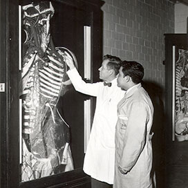
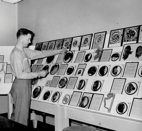
 Henry H. Hoffman, M.D., and student Manuel De Los Santos view a papier mache anatomical model, circa 1950. Hoffman was a professor in the anatomy department after 1957. While computer-based learning modules and high-tech gadgetry are central to medical training today, nothing compares to a physical object you can hold and inspect closely to arouse curiousity and a passion for discovery. Presented here are a few training tools and methods from the School of Medicine's past that still inspire fascination.
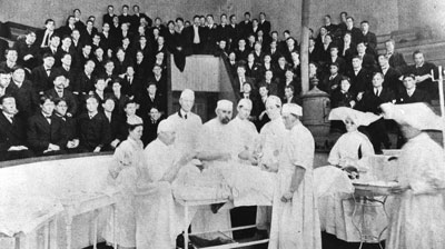 Surgical demonstration for students of the Medical College of Alabama in Mobile, circa 1904. After 1897 the Mobile school was (nominally) a department of the University of Alabama but retained its own governing board. In 1907 the Mobile board was dissolved and the University of Alabama became the governing entity for the medical school.
 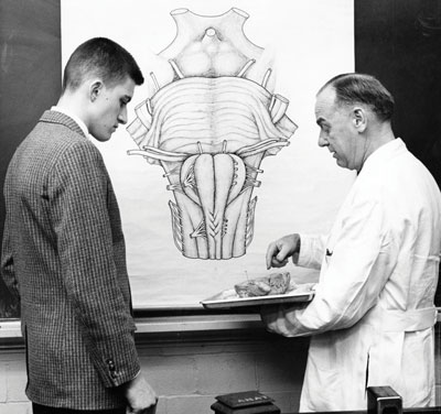 Dr. Thomas E. Hunt (right) and a student review a specimen, circa 1961. Hunt was an anatomy professor in Tuscaloosa from 1929 until 1945, and in Birmingham from 1945 until 1967.
 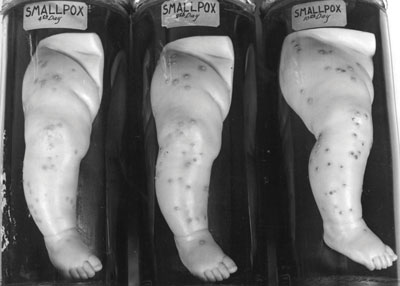 Stages of smallpox as portrayed in wax models in the Nott Specimen collection, circa 1950s. These 19th century models are currently located in the Alabama Museum of the Health Sciences, UAB Libraries.
  Edward L. Ramsey with the Boyd Pathological Specimens, circa 1959. Ramsey was with the autopsy pathology laboratory for over three decades. The Boyd Specimens is one of the collections housed in the Alabama Museum of the Health Sciences, UAB Libraries.
By Tim L. Pennycuff
Images courtesy of UAB Archives