 In the mouse brain, two neural pathways were discovered: The first is active during motivation; the second is active only at the termination of motivation. In humans, these pathways could underlie motivational dysfunctions present in various psychiatric conditions.Hunger can drive a motivational state that leads an animal to a successful pursuit of a goal — foraging for and finding food.
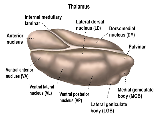
In a highly novel study published in Current Biology, researchers at the University of Alabama at Birmingham and the National Institute of Mental Health, or NIMH, describe how two major neuronal subpopulations in a part of the brain’s thalamus called the paraventricular nucleus participate in the dynamic regulation of goal pursuits. This research provides insight into the mechanisms by which the brain tracks motivational states to shape instrumental actions.
For the study, mice first had to be trained in a foraging-like behavior, using a long, hallway-like enclosure that had a trigger zone at one end and a reward zone at the other end, more than 4 feet distant.
Mice learned to wait in a trigger zone for two seconds, until a beep triggered initiation of their foraging-like behavioral task. A mouse could then move forward at its own pace to the reward zone to receive a small gulp of strawberry-flavored Ensure. To terminate the trial, the mice needed to leave the reward zone and return to the trigger area, to wait for another beep. Mice learned quickly and were highly engaged, as shown by completing a large volume of trials during training.
The researchers then used optical photometry and the calcium sensor GCaMP to continuously monitor activity of two major neuronal subpopulations of the paraventricular nucleus, or PVT, during the reward approach from the trigger zone to the reward zone, and during the trial termination from the reward zone back to the trigger zone after a taste of strawberry-flavored food. The experiments involve inserting an optical fiber into the brain just about the PVT to measure calcium release, a signal of neural activity.
The two subpopulations in the paraventricular nucleus are identified by presence or absence of the dopamine D2 receptor, noted as either PVTD2(+) or PVTD2(–), respectively. Dopamine is a neurotransmitter that allows neurons to communicate with each other.
“We discovered that PVTD2(+) and PVTD2(–) neurons encode the execution and termination of goal-oriented actions, respectively,” said Sofia Beas, Ph.D., assistant professor in the UAB Department of Neurobiology and a co-corresponding author of the study. “Furthermore, activity in the PVTD2(+) neuronal population mirrored motivation parameters such as vigor and satiety.”
Specifically, the PVTD2(+) neurons showed increased activity during the reward approach and decreased activity during trial termination. Conversely, PVTD2(–) neurons showed decreased activity during the reward approach and increased activity during trial termination.
“This is novel because people didn’t know there was diversity within the PVT neurons,” Beas said. “Contrary to decades of belief that the PVT is homogeneous, we found that, even though they are the same types of cells (both release the same neurotransmitter, glutamate), PVTD2(+) and PVTD2(–) neurons are doing very different jobs. Additionally, the findings from our study are highly significant as they help interpret contradictory and confusing findings in the literature regarding PVT’s function.”
 Sofia Beas, Ph.D.,
Photo credit: Sofia BeasFor a long time, the thalamic areas such as the PVT had been considered just a relay station in the brain. Researchers now realize, Beas says, that the PVT instead processes information, translating hypothalamic-derived needs states into motivational signals via projections of axons — including the PVTD2(+) and PVTD2(–) axons — to the nucleus accumbens, or NAc. The NAc has a critical role in the learning and execution of goal-oriented behaviors. An axon is a long cable-like extension from a neuron cell body that transfers the neuron’s signal to another neuron.
Researchers showed that these changes in neuron activity at the PVT were transmitted to the NAc by measuring neural activity with an optical fiber inserted where the terminals of the PVT axons reach the NAc neurons. The activity dynamics at the PVT-NAc terminals largely mirrored the activity dynamics the researchers saw at the PVT neurons — namely increased neuron activity signal of PVTD2(+) during reward approach and increased neuron activity of PVTD2(–) during trial termination.
“Collectively, our findings strongly suggest that motivation-related features and the encoding of goal-oriented actions of posterior PVTD2(+) and PVTD2(-) neurons are being relayed to the NAc through their respective terminals,” Beas said.
During each mouse recording session, the researchers recorded eight to 10 data samples per second, resulting in a very big dataset. In addition, these types of recordings are subject to many potential confounding variables. As such, the analysis of this data was another novel aspect of this study, through use of a new and robust statistical framework based on Functional Linear Mixed Modeling that both account for the variability of the recordings and can explore the relationships between the changes of photometry signals over time and various co-variates of the reward task, such as how quickly mice performed a trial, or how the hunger levels of the animals can influence the signal.
One example of how researchers correlated motivation with task performance was separating the trial times into “fast” groups, two to three seconds to the reward zone from the trigger zone, and “slow” groups, nine to 11 seconds to the reward zone.
“Our analyses showed that reward approach was associated with higher calcium signal ramps in PVTD2(+) neurons during fast compared to slow trials,” Beas said. “Moreover, we found a correlation between signal and both latency and velocity parameters. Importantly, no changes in posterior PVTD2(+) neuron activity were observed when mice were not engaged in the task, as in the cases where mice were roaming around the enclosure but not actively performing trials. Altogether, our findings suggest that posterior PVTD2(+) neuron activity increases during reward-seeking and is shaped by motivation.”
Deficits in motivation are associated with psychiatric conditions like substance abuse, binge eating and the inability to feel pleasure in depression. A deeper understanding of the neural basis of motivated behavior may reveal specific neuronal pathways involved in motivation and how they interact. This could lead to new therapeutic targets to restore healthy motivational processes in patients.
Co-authors with Beas in the study, “Dissociable encoding of motivated behavior by parallel thalamo-striatal projections,” are Isbah Khan, Claire Gao, Gabriel Loewinger, Emma Macdonald, Alison Bashford, Shakira Rodriguez-Gonzalez, Francisco Pereira and Mario Penzo, NIMH, Bethesda, Maryland. Beas was a post-doctoral fellow at the NIMH before moving to UAB last year.
Support came from National Institutes of Health award K99/R00 MH126429, a NARSAD Young Investigator Award by the Brain and Behavior Research Foundation, and NIMH Intramural Research Program award 1ZIAMH002950.
At UAB, Neurobiology is a department in the Marnix E. Heersink School of Medicine.