 Three years ago today, UAB Hospital made history by opening Alabama's first intraoperative MRI (iMRI) suite.
On Feb. 8, 2022, the University of Alabama at Birmingham Department of Neurosurgery James Garber Galbraith Endowed Chair of Neurosurgery James Markert, M.D., MPH, performed the suite’s first procedure—a craniotomy to remove a brain tumor.
The state-of-the-art facility, which remains one of only a few in the mid-South region encompassing Kentucky, Tennessee, Arkansas, Louisiana, Georgia and Alabama, has transformed surgical precision and patient outcomes.
A vision realized
“This suite was sort of a vision that I had for many years,” said Markert. “The intraoperative MRI allows us to view brain images taken while the surgery is underway to confirm that we have resected as much of the tumor as possible while leaving healthy brain tissue intact.”
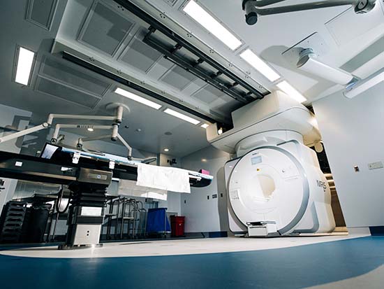
The iMRI suite at UAB Hospital employs the IMRIS Hybrid Operating Suite, which consists of two operating rooms with the MRI scanner positioned in a third room between them. The MRI is mounted on a specialized track system, allowing it to move into an operating room in just 90 seconds. Once in position, the machine slides over the surgical table, providing updated high-resolution imaging within minutes.
For patients undergoing neurosurgical procedures such as craniotomies or deep brain stimulation (DBS), this technology represents a major advancement in care.
 James Markert, M.D., MPH
Brain tumors, particularly gliomas, often have indistinct boundaries, making complete resection challenging. The ability to obtain updated MRI scans mid-surgery gives surgeons critical insights that were previously unavailable.
“The boundary between a brain tumor such as a glioma and the healthy brain tissue surrounding it is not necessarily well-defined,” Markert said. “Gliomas invade healthy brain cells by sending tendrils of tumor cells out from the primary tumor. The MRI helps a surgeon determine whether they have captured all of the tumor cells. We can recalculate as we operate.”
Enhancing patient outcomes
iMRI technology is particularly beneficial for conditions requiring a delicate and conservative surgical approach, such as epilepsy and movement disorders like Parkinson’s disease. DBS procedures, for example, rely on millimeter-level precision, making real-time imaging an invaluable tool.
In addition to brain tumor cases, the iMRI suite is also used for laser interstitial thermal therapy (LITT), a minimally invasive, MRI-guided laser procedure used to destroy brain tissue.
“The value of an intraoperative MRI is that we can look at brain images taken while the surgery is underway to confirm that we have resected as much of the tumor as possible while leaving healthy brain tissue intact,” said Markert. “Instead of relying on imaging that was taken hours or even days before, we are able to see the structures in the brain in real time, giving us the most complete and accurate view of the surgery.”
Since its inception in 2022, UAB Hospital’s iMRI suite has set a new standard for neurosurgical care in Alabama. By allowing surgeons to pause mid-procedure, obtain updated scans and adjust their approach in real time, this technology is improving patient safety, increasing surgical success rates and reinforcing UAB’s position as a leader in patient care and innovation.