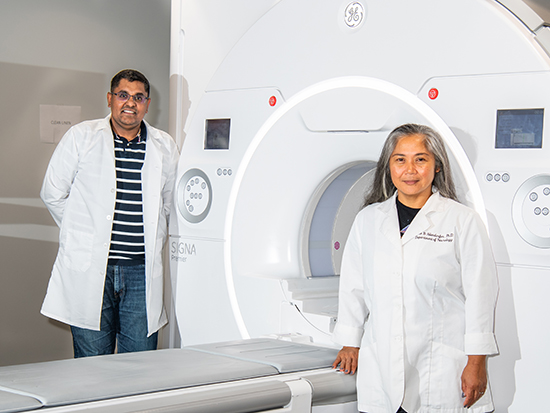
 Virendra Mishra, Ph.D., and Jane Allendorfer, Ph.D., with the GE PREMIER 3.0T whole-body MRI system at UAB Highlands, one of four scanners under the Research MRI Core.
When UAB investigators are interested in carrying out state-of-the-art experiments and analyses to examine brain and body anatomy and function, they come to the Research MRI Core, one of 15 core facilities in the UAB Institutional Research Core Program.
“We have four scanners under our core,” said core co-director Virendra Mishra, Ph.D., associate professor in the Department of Radiology. “The most widely used is the Prisma scanner at UAB Highlands.” This Siemens MAGNETOM is a powerful, 3.0 Tesla, state-of-the-art model. “Very few universities have this scanner,” Mishra said.
The higher the gradient strength of an MR scanner, measured in milli-Tesla/meter, “the faster we can go,” Mishra said. Because errors introduced by human movement are the major source of noise in an MR image, “when you can complete the scan quickly, you can not only go to a higher resolution but also add more exams during a fixed allotted time,” he said. “It is much faster and cleaner.” This is particularly helpful for researchers interested in the connectivity of the brain. “We want to image the neurons firing, but conventional scanners can’t go fast enough,” Mishra said.
New additions
The latest additions to the core are a GE Premier 3.0 Tesla wide bore scanner at UAB Highlands, a GE SIGNA PET/MR 3.0 Tesla model at the Advanced Imaging Facility in the Wallace Tumor Institute and, at the opposite end of the power range, a 0.55 Tesla Siemens Freemax at UAB Highlands. “People who have implants in their bodies cannot be scanned at 3 Tesla,” Mishra said. “But there is a real need to see MR imaging of these patients.” Typically, clinicians resort to CT, ultrasound and X-rays, “but MR cannot be fully replaced by other modalities,” he said. The lower-field 0.55 Tesla Freemax “can collect images at reasonable quality for these uses, and we are trying to revise studies to see how we can use it for research,” Mishra added.
The most common research use for the MR scanners is for brain studies, says Research MRI Core co-director Jane Allendorfer, Ph.D., associate professor in the Department of Neurology. “But people also use them for imaging the liver, cancer, lung disease, the pancreas, and a significant number of other uses,” she said.
In addition to the scanners themselves and the expertise in using them, the Research MRI Core provides advice on the best protocols to use for individual experiments and research questions. “If researchers don’t know, we help them find solutions on the technical side,” Allendorfer explained. “We don’t change the investigator’s aims, but we try to help them get the scanner set up in the best way.”
The core also offers additional equipment for functional MRI studies, including button boxes for participant responses while being scanned, headphones, and reflective screens for watching images or videos.
Regular quality assurance checks
Another important benefit of working with the Research MRI Core is its regular quality assurance checks to ensure that results are reproducible. This is crucial when investigators are part of multi-site studies, Allendorfer says. “Signal to noise is something that people are very concerned about with MRI,” she said. “We do weekly QA scans on phantoms, so we have the data tracked over time. That way, if an investigator sees something they were not expecting, we can go back to our QA scans for the week and say, ‘We saw those, too’ or let them know that this is more likely to be a real phenomenon they are seeing in their images.”
Investigators typically fall into two categories: those who have extensive MRI training, and those with well-established research in a disease or condition who want to add MRI to their studies. “Many of them focus on the brain’s structure and function and its metabolites, but we have the capability to image the whole body,” Allendorfer said. Researchers in the UAB Nutrition Obesity Research Center, for example, use MR to study the liver and pancreas, she notes. MR can also be used to study heart functioning and to image the diffusion of water through various muscles and tissue types.
Allendorfer’s own research is with epilepsy patients before and after an intervention consisting of endurance and resistance exercise training. “I have a pilot trial now studying endurance training in temporal lobe epilepsy,” she said. Patients with this form of epilepsy tend to have extensive memory deficits. “We’re looking at how exercise can mitigate some of these deficits.”
Coming soon: Mock scanner
An eagerly awaited new piece of the Research MRI Core offerings is a mock scanner that can simulate the sights, sounds and feelings of being in an MRI scanner without any imaging taking place. “A lot of people, including my mother and brother, are scared to go into this closed system of an MRI,” Mishra said. “They will come to get scanned and then back out. With the mock scanner, they will be able to get a feel for it, understand how the scan will be conducted and how long it takes, what it sounds like, all of that.” This acclimatization will be particularly useful for research studies, where time and expense is accrued when participants find themselves unable to go into the scanner.
The machine will closely mimic the Prisma MR scanner at UAB Highlands and be installed nearby. Other planned purchases include an EEG/fMRI system for simultaneous scanning capability, which will be very helpful for brain studies, Mishra says.
Working with the Research MRI Core
The best way to begin working with the core is to email
It is always best if investigators reach out as soon as possible, Allendorfer adds. “When they reach out to us first before they propose a study, we can suggest what can be done and what cannot be done,” she said. “We can also let them know the fees for the scans so they can budget accordingly,” especially if the imaging needs to be done at night or has other requirements that will add additional costs.
The core does not provide data analysis support, Mishra says, mainly because the correct solution is very dependent on the individual study questions. “There are a significant number of software platforms in this field, and we can guide investigators to resources that will help them identify the best for their research,” he said.
Learn more about the Research MRI Core at www.uab.edu/cores/ircp/rmric.
New users: Start here
The best way to begin working with the core is to email