Over the past several years, word has spread in the neuroscience research community: UAB is hiring.
The first two rounds of the Strategic Neurosciences Recruitment initiative at the Heersink School of Medicine began pre-pandemic, with 28 new faculty hired across 11 UAB departments. The third round, which launched in summer 2023, is seeking up to 20 more faculty and included a full-page ad on the back cover of the journal Nature. By mid-May 2024, the call had brought more than 170 applications, says Jeremy Day, Ph.D., director of UAB’s Comprehensive Neuroscience Center, who is contributing to the recruiting effort. The initiative is co-chaired by David Standaert, M.D., Ph.D., chair of the Department of Neurology and director of the NIH-funded Alabama Morris K. Udall Center of Excellence in Parkinson’s Disease Research, and Erik Roberson, M.D., Ph.D., director of the UAB Alzheimer’s Disease Center.
“These are researchers with competitive offers from our peer institutions and we convinced them to come here,” said Day, an associate professor in the Department of Neurobiology who also leads the School of Medicine’s joint task force on brain health and disease across the lifespan. “Trying to recruit at this level, with the quality of the candidates that we have, is a tough game. I’m really happy with the success that we have had.”
“I want to recognize the tremendous leadership of Senior Vice Dean Dr. Tika Benveniste, who has for many years spearheaded our recruitment efforts for key research areas in the UAB Heersink School of Medicine through our Strategic Recruitment Office and the Office of Research," said Anupam Agarwal, M.D., senior vice president for Medicine and dean of the Heersink School of Medicine. "This approach that she initiated for our research faculty recruitment has been transformative and allowed us to grow by hiring exceptional faculty across the school and specifically in the important area of neurosciences. We look forward to Dr Benveniste’s continued leadership as we focus on and prioritize our efforts in support of the Growth With Purpose initiative.”
As Agarwal notes, this recruitment success aligns with UAB’s Research Strategic Initiative: Growth with Purpose, announced in summer 2023, which sets out a road map for future research growth. They already have raised UAB’s elite neuroscience research community to a new level of excellence. Over the past decade, the university’s brain-related research funding from the National Institutes of Health has more than doubled.
That growth has tracked with a nationwide realization that brain health is one of the biggest challenges of modern times. The country’s aging population faces a surge in neurodegenerative conditions and simultaneous crises in addiction, mental health disorders, chronic pain, and intellectual and neurodevelopmental disabilities. Alabama’s population is older and poorer than the national average, with less access to health care and more health inequity, all of which are risk factors for brain health.
But UAB is uniquely poised to address these crises, Day points out. “Our goals are to understand brain disease conditions, identify the factors that are disrupted in brain disease states, harness neurotechnology to treat brain diseases, and transform care of brain-related diseases in Alabama.”
New recruits have already set up labs studying feeding-related fear signaling, skeletal muscle pathways that can alter the rate of brain aging, genetic timers that control neural development, sex-related differences in psychiatric illnesses, the effects of brain training on dementia risk, and the role of gut-related microbiota on epilepsy and Alzheimer’s disease, among many other research questions.
How do you attract scientists who are doing groundbreaking work? As the Nature ad highlights, UAB has size on its side: the Comprehensive Neuroscience Center has a network of more than 500 faculty, researchers, clinicians, staff, and trainees from 32 departments and nine schools. UAB has more than 730,000 square feet of research space and recently broke ground on the new Biomedical Research and Psychology Building, which will house many neuroscientists. Later this year, the new Altec/Styslinger Genomic Medicine and Data Sciences Building will open. “It will house data scientists who are crucial for unravelling the mysteries of the brain with large-scale datasets,” Day said. UAB also is home to 15 campus core facilities with state-of-the-art equipment and expertise to support cutting-edge research. And it is a national leader in patient care; UAB Hospital is one of the largest hospitals in the United States.
The Strategic Neuroscience Recruitment effort is not restricted to faculty, Day adds. The innovative Brain-PRIME postdoctoral fellowship program offers a two-year award, starting salary of $70,000, a place in the neuroscience lab of the recipient’s choice, advanced mentoring, and $5,000 in career development funds. In its inaugural year, the program received 27 applications, extended six offers and had five acceptances. In January 2024, the NeuroScholars award program launched to help recruit top graduate student candidates to the neuroscience theme of UAB’s Graduate Biomedical Sciences training program; it provides $10,000 in cash and scientific discretionary funds. “We are actively recruiting talented trainees at all levels,” Day said.
Meet five new faculty recruits who explain their work and why they decided to come to Birmingham to move their research forward.
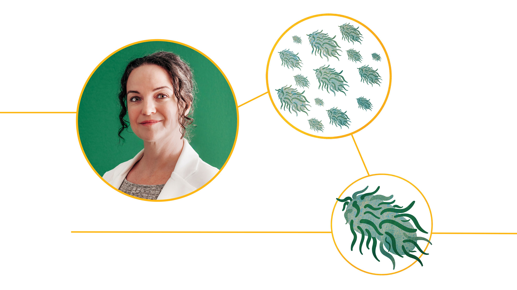
Bugs as drugs
Angela Carter, Ph.D.
Assistant Professor, Surgery
Many microbes attack the body, but not all. Some are symbionts that help defend us against other invaders, provide metabolic help or even assist with nutrients we could not get any other way. And others have shadowy roles—they are not attacking the body themselves, but they seem to help others do so.
“We know that microbes are present in many different niches in the body and they are naturally altering our physiology,” Carter said. “My lab is trying to understand how microbes do this so that we can leverage those molecular mechanisms toward the treatment of human disease. In addition, we are taking a synthetic biology approach, to engineer bacteria to produce therapeutic molecules, so that we can directly capitalize on these relationships between microbes and hosts.”
Carter’s lab is studying ways to use probiotics to boost helpful bacteria, examining prebiotics that induce microbes to produce useful byproducts, co-opting bacteria-targeting viruses, known as phages, to eradicate disease-promoting bacteria populations, as well as genetically designing bacteria to make beneficial products.
In April 2024, Carter became the first UAB faculty member to receive a Discovery Boost Grant from the American Cancer Society. Her project seeks to create microbes capable of producing therapeutic components within tumors. Specifically, she is engineering a strain of Salmonella bacteria to produce monoamine oxidase (which degrades serotonin) and insulin-degrading enzyme (which does just what it says) to counteract the toxic over-production of these hormones by neuroendocrine tumors.
Carter started out in the field studying Fusobacterium nucleatum, which typically resides in the mouth. However, Fusobacteria also are found at abnormally high rates in the gut microbiomes of people using amphetamine drugs, including the dangerous stimulant crystal meth. There is a connection: Fusobacteria secrete a short-chain fatty acid called butyrate, which crosses the blood-brain barrier and plays a role in maintaining the extracellular dopamine release that is responsible for amphetamines’ stimulant effect. But do amphetamines help Fusobacteria to flourish, or is it the other way around? Carter and UAB’s Aurelio Galli, Ph.D., Distinguished Professor in the Division of Gastrointestinal Surgery, “have found that it is not a unidirectional relationship—it is a feed-forward cycle,” Carter said. “Amphetamines actively increase the abundance of Fusobacteria, and Fusobacteria then target the dopamine transporter in brain cells to enhance the function of amphetamines.”
Amphetamines also act on other members of the microbial community to boost production of enzymes known as glucosyl transferases, which help Fusobacteria produce the protective biofilm barriers that make them resistant to environmental stressors such as antibiotics. Carter and Galli are supported by two NIH grants totaling $3.1 million, awarded last fall and this spring, that focus on understanding exactly how Fusobacteria affect substance use disorders and exploring ways to disrupt biofilms and crush Fusobacteria populations.
Why UAB? Carter originally came to UAB as a postdoc when her lab’s principal investigator was recruited to Birmingham. “The main reason I stayed, after my postdoc finished, was the great collaborative network I found here,” Carter said. “It all started working on substance use disorders with Dr. Galli, who has been an amazingly supportive colleague and mentor. Now my lab has branched out into studying how microbes impact multiple different diseases, and I continue to find engaging collaborators all throughout UAB.”
<div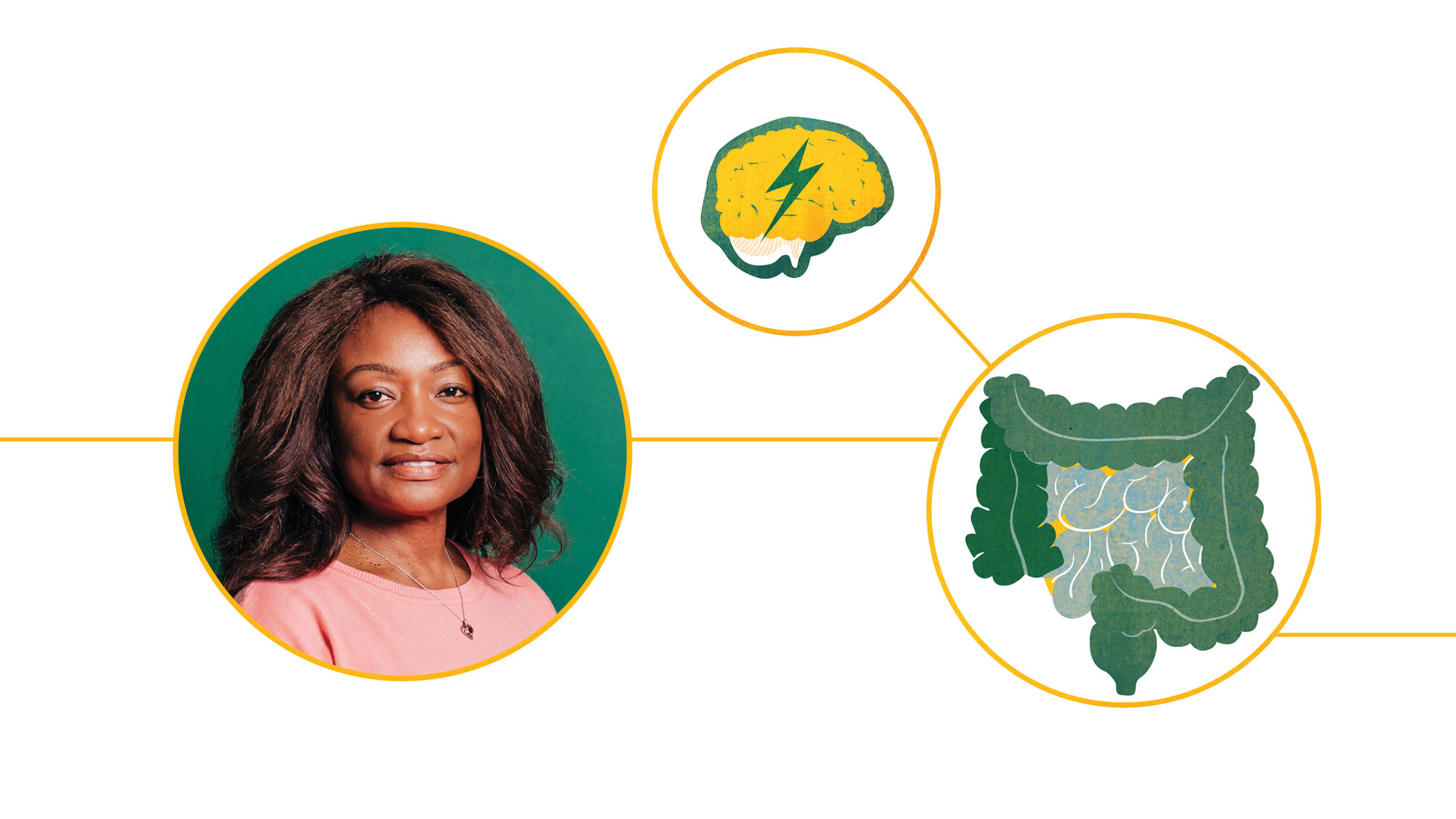
Stopping seizures through the gut
Susan L. Campbell, Ph.D.
Associate Professor, Cell, Developmental & Integrative Biology
Since the early 1930s, doctors have known that the high-fat, low-carb ketogenic diet can help reduce seizures. But the mechanisms were unclear, until a 2018 paper demonstrated that the diet induced changes in the gut microbiota that conferred seizure protection in models of refractory epilepsy.
By the time the paper was published, Campbell had already begun to work on this area in other animal models. “There were other pieces of evidence showing a difference in the composition of the gut microbiota between epilepsy patients who respond to treatment and those patients with drug-resistant epilepsy,” Campbell said.
Inspired by this evidence, Campbell’s lab “really started to ask questions about what the gut microbiome can tell us about seizure genesis,” she said. Using a mouse model of virus infection-induced seizures, her team has shown that specific microbes were present in the seizure-free animals (and reduced in animals that had seizures). “And we were able to take it a step further, which not all studies do, and look at the functions of those specific microbes that were underrepresented in seizure animals,” Campbell said. In a 2021 paper, her lab showed that virus-infected mice who went on to have seizures showed a loss in several bacteria that produce a gut metabolite called S-equol—and that S-equol reduced the hyperexcitability seen in neurons from animals with seizures. “We were very excited about that finding, because it gives us a potential target to assess,” Campbell said. She now has a large NIH grant “to dissect the full mechanism.”
Another area of interest in Campbell’s lab is studying how existing anti-seizure medications change the gut microbiome “and if those interactions impact the efficacy of the medication,” she said. Also, her lab recently obtained NIH funding for a new study examining the effects of vaping nicotine and flavorings on seizure susceptibility and the function of brain cells.
Why UAB? Campbell first came to Birmingham in 1999 for a Ph.D. in the lab of Professor John Hablitz, Ph.D., who studies epilepsy at the functional level. Campbell had become fascinated with brain function—and epilepsy in particular—as an undergraduate McNair Scholar at the State University of New York at Binghamton. After Campbell completed her doctorate at UAB, she stayed on to do a postdoc in the lab of Harald Sontheimer, Ph.D., then a professor and director of UAB’s Civitan International Research Center. Campbell herself launched the Civitan International Research Center Research Club, which gave her the opportunity to inform Civitan members across the country and internationally about ongoing work at the center. After Sontheimer moved to Virginia Tech in 2015, Campbell went there and then started a faculty position in 2018. “But as the research funding and timing in terms of life made me ready to make a move, I knew that UAB would always be at the top of my list when I was ready to move,” she said. “The research environment at UAB is unparalleled. All you have to do is come here to see that.”
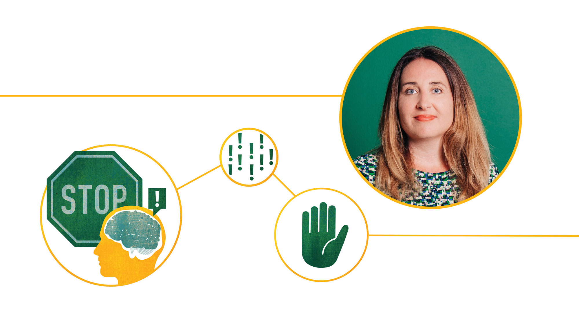
Blocking bad memories
Jamie Peters, Ph.D.
Associate Professor, Neurobiology
Peters’ lab is looking for new treatment interventions for addictive drugs and fear-related disorders. She has a particular focus on a region of the prefrontal cortex that in humans is known as Brodmann area 25. “It seems to be very important for self-control, and deficits in self-control are a hallmark characteristic of people suffering from substance use disorders,” Peters said. “There has been a lot of work to support the involvement of the prefrontal cortex in all sorts of behaviors and inhibiting emotional responses when they are inappropriate.” Addiction, like post-traumatic stress disorder, “is an emotional memory disorder,” Peters explained. “In one case it is a ‘good,’ rewarding memory and in the other it is a bad, traumatic memory, but in both it is an emotional memory.”
In animal models, Peters’ lab has been able to block these emotional memories in two ways: with designer receptors or with light-sensitive channels. “Chemogenetics and optogenetics have revolutionized the field of systems neuroscience,” she said. Using a specially prepared virus with a modified gene payload, Peters can induce neurons in the prefrontal cortex to express “dummy” receptors that are only activated by designer drugs such as J60 hydrochloride. Giving the drug inhibits the neurons in these memory circuits, stopping them from firing. The result: the animals relapse at lower rates when confronted by drug cues.
Peters’ lab also uses optogenetic techniques—delivering genes via direct injection or modified viruses that can then be turned on and off through bursts of light. “We have been able to figure out a lot of the specific neural circuits that are able to control different types of behavior, including relapse behavior,” she said.
A new and exciting area of investigation for the lab, Peters says, involves testing the effects of psychoplastogens. These are lab-made compounds that retain the therapeutic value of psychedelic drugs such as psilocybin without the hallucinogenic effects. Peters is collaborating with David Olson, Ph.D., a medicinal chemist at the University of California-Davis. They have demonstrated that the lab-created drug tabernanthalog “has therapeutic effects in preclinical models of opioid-seeking behavior,” Peters said.
Why UAB? The collaborative potential at UAB “was a huge draw” in bringing her to Birmingham, Peters says. She had already begun a collaboration with Day while she was still at the University of Colorado-Denver to understand “what is genetically different about these cells in the prefrontal cortex after drug use,” Peters said. “Jeremy is doing elegant RNA sequencing work; his work at the genetic level and my work at the systems level allows us to really come at the problem of drug addiction from interesting angles.”
Where is language in the brain?
Matthew Nelson, Ph.D.
Assistant Professor, Neurosurgery
In graduate school, Nelson began his career as an intracranial electrophysiologist—mapping electrical activity in the brain—by studying intracranial messaging in non-human primates. At the time, “there was this view that animal electrophysiology was more serious,” he said.
But as he watched a colleague at CalTech develop intriguing results in humans—working with patients who had electrodes implanted in their brains prior to epilepsy surgery—Nelson decided to make the switch himself. “We can now take the best experiments that have been developed in animals and do them in humans,” he said. “We have these unique opportunities to record in humans and I thought we should study something we can only study in humans. And that led me to language.”
As a postdoc, Nelson worked in the lab of noted cognitive neuroscientist Stanislas Dehaene, Ph.D., at the Neurospin Research Center in France. Nelson was first author on a much-discussed 2017 paper with Dehaene that recorded brain activity in the left hemisphere language network while participants read a series of sentences. The results of those experiments provided evidence validating the linguistic theory that humans comprehend sentences by grouping them in a “tree-like hierarchy of nested phrases,” rather than parsing them one word at a time, as another theory held.
In a second postdoc at Northwestern University, Nelson worked with a neurosurgeon and the renowned cognitive neurologist Marsel Mesulam, M.D., on a series of studies in patients with primary progressive aphasia. In this rare neurodegenerative disorder, patients have difficulty understanding words, finding the right word or expressing themselves.
At UAB, Nelson has established collaborations with epileptologists and neurosurgeons in the UAB Epilepsy Center. Patients with treatment-resistant seizures often undergo intracranial EEG monitoring to locate the source of the seizures so that surgeons can remove the faulty tissue. “The neurosurgeons place the electrodes and we make use of the opportunity to collect the best neural data possible,” Nelson said. “This is an incredible opportunity to better map language in the brain.”
Nelson also has the chance to study patients after entire brain regions, such as the anterior temporal lobe, are removed during seizure surgeries. His work is adding to evidence that some of these regions play a role in language processing and generation, expanding the boundaries of the brain’s language network. Better mapping of language areas will help surgeons avoid these areas during brain resections, Nelson explained. “Preserving language is critical to patients’ quality of life.”
Why UAB? In addition to his collaborations with UAB epileptologists, Nelson was attracted by UAB’s equipment for non-invasive study of brain circuits, including magnetoencephalography and transcranial magnetic stimulation machines. His work also has clear application to brain-machine interfaces, which are the focus of UAB’s latest university-wide interdisciplinary research center, the Center for Neuroengineering and Brain-Computer Interfaces.
Brain changes that matter
Virendra Mishra, Ph.D.
Associate Professor, Radiology
Repetitive head trauma, of the type experienced by boxers, football players and other athletes, is a well-known contributor to chronic traumatic encephalopathy, or CTE. But not everyone who takes the football field will develop CTE, just as many older adults never experience the horrors of dementia.
Mishra specializes in combining MRI images with novel computational tools, including machine learning and deep learning algorithms, to predict cognitive decline in Parkinson’s disease and in patients with repeated head impacts. At the Cleveland Clinic, Nevada in Las Vegas, Mishra developed a model trained on data from nearly 300 boxers; it identified seven MRI-derived features that predict which fighters will go on to develop CTE with 75 percent accuracy.
The collaboration, which has continued after Mishra was recruited to UAB, aims to identify boxers at highest risk of long-term damage. “That would allow them to make a lifestyle change and define a return to play guideline so that their brains can adjust,” Mishra said.
Mishra’s predictive models are also trained on advanced MRI imaging of patients with Parkinson’s disease, combined with demographic information, genetic signatures, and biomarkers from cerebrospinal fluid.
But before MRI scans can be used for widespread diagnosis, there is another issue to be solved: reproducibility. “If you get scanned in one machine and then later come back and get scanned in a second machine, the data will not be equal,” Mishra said. The issue is with the “noise”—artifacts that are an unavoidable result of the imaging process and can vary based on the manufacturer of the scanner and the settings (including spatial and angular resolutions) of the machines. That makes detailed comparisons challenging. “How do I compare a patient’s brain today with the patient’s brain before they had Parkinson’s, even if I have that image?” Mishra said. “Our lab is working to define protocols that are consistent and reproducible and develop software to remove the noise.”
Why UAB? One major draw for Mishra was the university’s Alabama Udall Center of Excellence in Parkinson’s Disease Research, which is focused on identifying and developing therapeutics for neuroprotection against Parkinson’s disease. “UAB is one of the few centers in the country with an NIH-funded Udall center and it is home to legends in the field such as Dr. David Standaert and Dr. Erik Roberson,” he said. “When Dr. Standaert called me for an interview, I couldn’t say no.” UAB’s Department of Radiology also has a state-of-the-art 3 Tesla Prisma MRI scanner and is one of the few that has a simultaneous PET-MRI scanner, he said. “The presence of the clinical population and the availability of top-of-the-shelf MRI scanners are ideal for my work on developing reproducible MRI protocols, testing the protocols and further designing predictive models based on those reproducible protocols on the patient population that has the most chances of early intervention to stop disease progression.” The university also has a pool of talented students for Mishra to recruit to his lab. “Access to students is essential for my work,” he said.
