


The Zeiss Lightsheet 7 for 3D imaging of live and cleared samples.
This microscope provides fast and gentle imaging of living model organisms, tissues, and organoids as well as fixed optically cleared specimens. Specimens can be up to 2 cm in size and at any refractive index between 1.33 and 1.58 compatible with most clearing solutions. Acquires lower resolution overview images or whole volume subcellular resolution.
High quantum efficiency detectors enable observations of the fastest processes at the lowest illumination light levels. The light sheet geometry illuminates only the parts of the sample you are imaging, greatly reducing photobleaching and phototoxicity. You'll get a real-life view of your samples without the adverse effects of excitation light on their biology.
FEATURES:
- Double side illumination objectives with single detection objective
- Environmental module with CO2, humidity, and temperature control
- Four imaging chambers for imaging live or fixed and cleared samples in broad range of sizes
- Two PCO Edge 4.2 cameras with 16bit sCMOS sensor, 2048x2048 pixel density, 5µs to 5s exposure time, and maximum frame rate of 40fps at full resolution.
- Four fluorescence imaging channels (blue, green, red, and far red).
- Advanced acquisition and analysis software operating of Lightsheet computer (Zeiss Zen Black- acquisition) and two high end workstations; WS1 (Zeiss Zen Blue, Arivis Vision4D), and WS2 (Imaris 10). All three computers are interconnected with 10Gbit fiber-optic allowing for fast transfer of data to either WS1 or WS2.
DETECTION OBJECTIVES:
Water Dipping Objectives:
- W Plan-Apo 20x/1.0 D, NA=1.0, WD=2.4mm - This objective is designed for dipping into physiologic salt solution. The correction collar is used to adjust small deviation of refractive index in the range of nD from 1.33 to 1.36.
- W Plan-Apo 10x/0.5, NA=0.5, WD=3.7mm - This objective is designed for dipping into physiologic salt solution.
Dry Objectives:
- EC Plan-Neofluar 5x/0.16, NA=0.16, nD=1.33-1.58 - This objective is designed for over the air imaging. The correction collar is used to correct refractive index in range of nD from 1.33 to 1.58.
- EC Plan-Neofluar 5x/0.16, NA=0.16, WD=18.5mm – This objective is designed for over the air imaging. For use with Translucence chamber (requires adaptor).
- Fluar 2.5/0.12, NA=0.12, WD=8.7mm - This objective is designed for over the air imaging. For use with Translucence chamber (requires adaptor).
Clear Dipping Objectives:
- Clr Plan-Neofluar 20x/1.0 Corr, NA=1, WD=6.4mm, nD=1.53 - Suitable for imaging samples processed with clearing methods with nd=1.53 +/- 0.05; for example U.Clear, Ce3D, Cubic Cancer.
- Objective Clr Plan-Neofluar 20x/1.0 Corr, NA=1, WD=5.6mm, nD=1.45 - Suitable for imaging samples processed with clearing methods with nd=1.45 +/- 0.05; for example FocusClear TM.
ILLUMINATION OBJECTIVES:
- LSFM 5x/0.1 foc - Pair of illumination objectives with correction collar for refractive index adjustment from nD of 1.33 to 1.58.
- LSFM 10x/0.2 foc - Pair of illumination objectives with correction collar for refractive index adjustment from nD of 1.33 to 1.58.
LASERS:
- 405nm
- 488nm
- 561nm
- 638nm
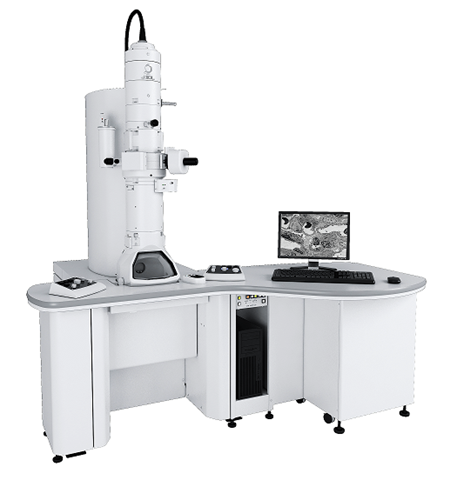
The JEOL 1400 High Contrast Transmission Electron Microscope is a state-of-the-art imaging system designed for high-resolution transmission electron microscopy (TEM). This microscope features advanced optics and imaging capabilities for biological, materials science, and nanotechnology applications. The JEOL 1400 TEM is an essential tool for researchers seeking high-resolution insights into the structure and composition of their samples, supported by specialists for optimal performance and operation.
Users benefit from intuitive software for image acquisition and analysis, streamlining workflows and enhancing productivity.
The JEOL 1400 TEM is equipped with a AMT-NanoSprint43L-MarkII Camera. With a remarkable resolution of 43 megapixels, this camera captures high-quality images with exceptional detail, making it an invaluable tool for researchers. Its cutting-edge sensor technology provides outstanding sensitivity and dynamic range, allowing for clear imaging of both bright and dark features in a wide variety of samples.
The user-friendly interface and comprehensive software package facilitate straightforward operation, making it easy for researchers to obtain and analyze images efficiently.
Features:
- The JEM-1400 can change the magnification smoothly from extremely low magnification(10X) to a maximum magnification of 1.5 million times without image rotation, and it is always possible to observe with high contrast.
- High-resolution imaging capability with a lattice resolution of .2 nm and a point resolution of 0.38 nm.
- Optimized for high contrast imaging for biological, low Z, and materials science applications
- AMT-NanoSprint43L-MarkII: AMT-NanoSprint43L-MarkII Camera, 43MP, low mount position on electron column
- +/- 70º tilt with support for tomography
- SerialEM software for montaging and tomography.
- LaB6 emitter standard
- Accelerating voltage range 20kV to 120 kV for optimal imaging.
- High-contrast imaging mode for low-contrast specimen visualization.
- Integrated automated stage for precise sample positioning.
- User-friendly interface and comprehensive software for efficient operation.
HRIF provides access, training, and support for state-of-the-art fluorescence and electron microscopes.
Follow the links below to explore what we have to offer! We are always available to assist you in selecting the best microscope or technique for your research!
Transmission Electron Microscopy
Digital Microscopy & Light Microscopy: Shelby Building Room 136D
 The HRIF contains several advanced, digital, fluorescence and light Microscopes which can be used to image cells and tissue. The Light microscopes are ideal for imaging cells and tissue that have histological stains. (I.e. Lung, Brain, Spleen, etc.)
Nikon Diaphot. Shelby 135B
This scope has fluorescence and brightfield capabilities with a wide range of magnifications from 2.5x to 100x. It’s primarily used for doing Brightfield, DIC, and Phase imaging. Notably, it can be used with the Q-Imaging Color camera for imaging histological stained sections and. The Diaphot Objectives have a long working distance and may be able to penetrate the plastic of certain Petri dishes that most other microscopes cannot, for that reason it is very useful for looking at live cells which have fluorescence.
Nikon SMZ-U Stereo/Dissection Scope. Shelby 135B
This microscope is great for imaging large slices of of lung, brain, and other large tissue samples that require magnification below 3x. It has the capability to do Fluorescence but is mostly use for Brightfield applications. In conjunction with the Q-Imaging High Res. Color camera it can image a variety of large scale samples for densitometry and quantitative purposes.
The Leica DRMB Fluorescent/Light Microscope (10x, 20x, 40x, 60x, Plan Apo Objectives) can be used for quick fluorescence viewing, pictures of light and fluorescence. Ideal for imaging H&E stains of cells and tissue in conjuction with the Q-Imaging Color Camera.


Sub-Micron Particle Imaging System (Exosomes)
The NS300 can visualize and measure particles in suspension in the size range 10-1000nm (depending on material) and addresses the needs of a wide variety of applications including protein aggregation, exosome and microvesicle research, drug delivery systems, and analysis of particles labeled with green fluorophores.
The NanoSight300 provides multi-faceted analysis that includes a motorized filter wheel, polarizer for analysis of bi-fringent particles, high sensitivity CMOS camera, temperature control and 488nm laser.
The top plate and laser module block are removable to enable easy cleaning and assembly. The top plate is interchangeable.