Case of the Week
- Details
Case History
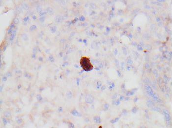
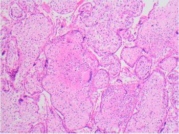
A 66-year-old female presented due to a cystic lesion of the pancreatic head approximately 5.6 cm. A Whipple procedure was performed and 5.7 cm-sized ill-defined multiloculated cystic mass involving the main pancreatic duct was identified by gross examination. Figures include H&Es, immunohistochemical stain (HepPar-1), and albumin mRNA in situ hybridization (ISH).
- Details
Case History
35-year-old male with excisional biopsy of left cervical lymph node.
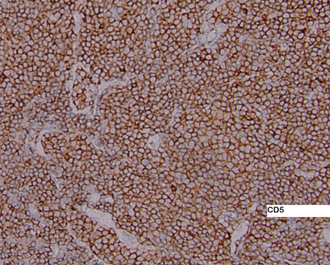
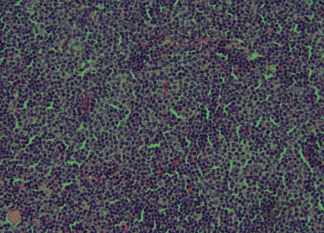
- Details
Case History
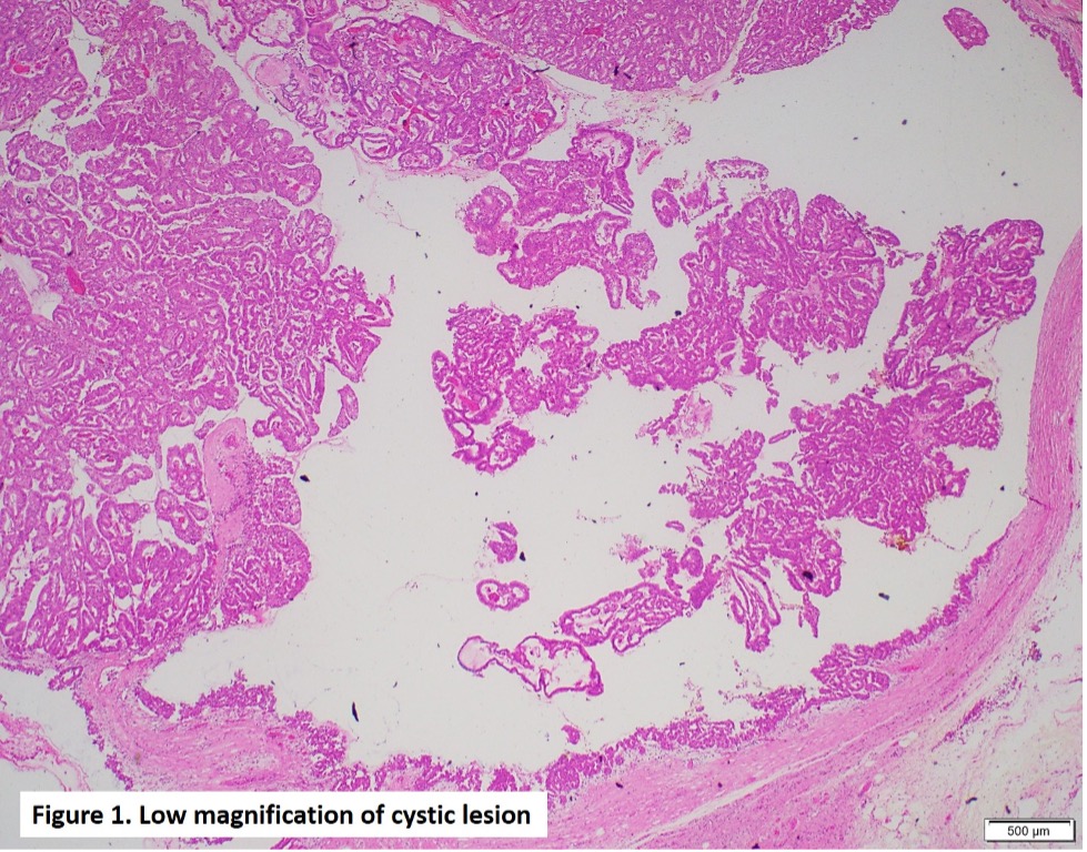
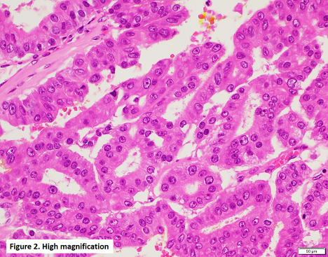
A 51-year-old female presented with extensive microcalcification in the left breast. The morphology of the partial mastectomy is shown in the figures. ER was positive in 50% of the lesional cells.

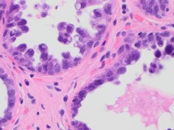
What is the diagnosis?
A. Apocrine ductal carcinoma in situ
B. Apocrine metaplasia
C. Cystic hypersecretory intraductal carcinoma
D. Secretory carcinoma
Correct Answer: C. Cystic hypersecretory intraductal carcinoma
Case contributed by: Xiao Huang, M.D., Ph.D., Assistant Professor, Anatomic Pathology, UAB Pathology
- Details
Case History
A 26-year-old female presents with a 11 cm unilateral ovarian mass and massive ascites. Immunohistochemical test for SMARCA4 / BRG1 shows loss of expression.


- Details
Case History
A 40-year man was brought to the ER with multiple injuries due to MVA. On arrival BP 60/35 mm Hg, HR 125 bpm. He transfused with 5 units of type O, Rh + whole blood over 25 minutes which improved BP to 90/50 mm Hg. After transfusing 5 more units BP dropped to 50/30 mm Hg, and the HR rises to 130 bpm. The electrocardiogram shows a QT interval of 470 msec. Central venous pressure increases from 10 to 22 cmH2O.
- Details
Case History
A 44-year-old female with a past medical history of both breast and thyroid cancer at age 32 (both appropriately treated) now presents with an acetabular mass and pathologic fracture.


