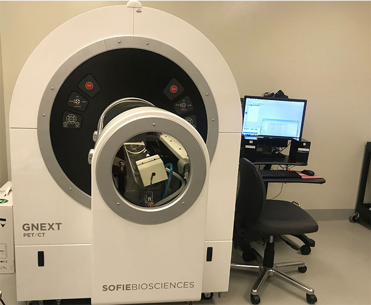
The PET/CT scanner uses imaging cameras, detectors, and radioactive tracers to create images based on the metabolic or functional activity of a cell. CT uses X-rays to provide detailed anatomical information, including the location, size and shape of lesions or tumors. PET/CT combines positron emission tomography (PET) with Computed tomography (CT) technology to aid in the diagnosis of cancer and in determining the extent to which cancer has spread.
The PET/CT was installed in 2017 and recently underwent a major upgrade adding a novel 2-D high throughout preclinical PET imaging system, the Beta-eye, that allows high-throughput dynamic analysis of novel radiopharmacetuicals to probe unique molecular processes in vivo in animal models longitudinally through time. This instrument can image mice and rats in dynamic or static images.
