Case History
56-year-old female with left floor of mouth erythematous lesion
What is the diagnosis:
- Melanoma
- Myoepithelioma
- Epithelioid Schwannoma
- Ectomesenchymal Chondromyxoid Tumor (ECMT)
The answer is “C” Epithelioid Schwannoma
Discussion
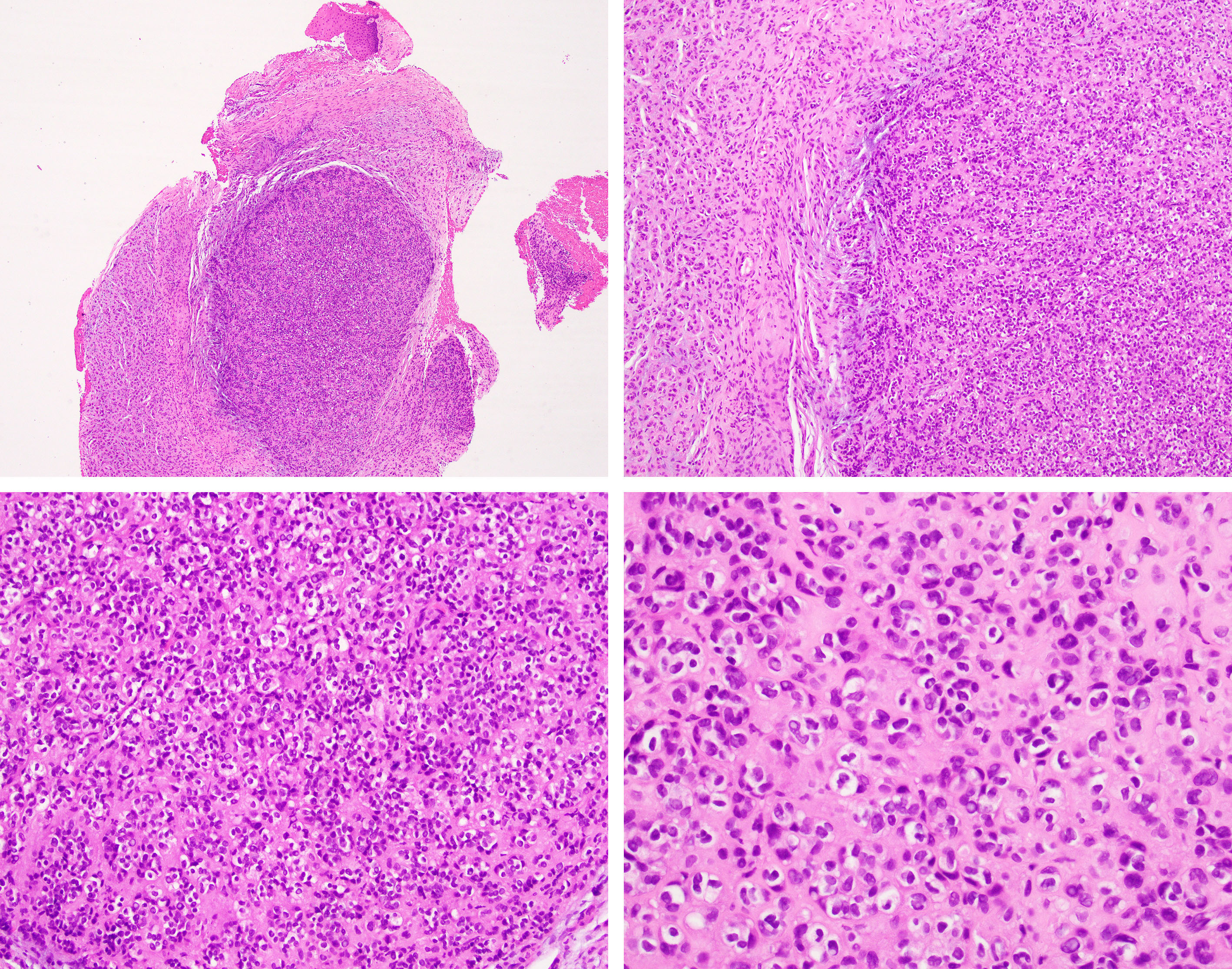
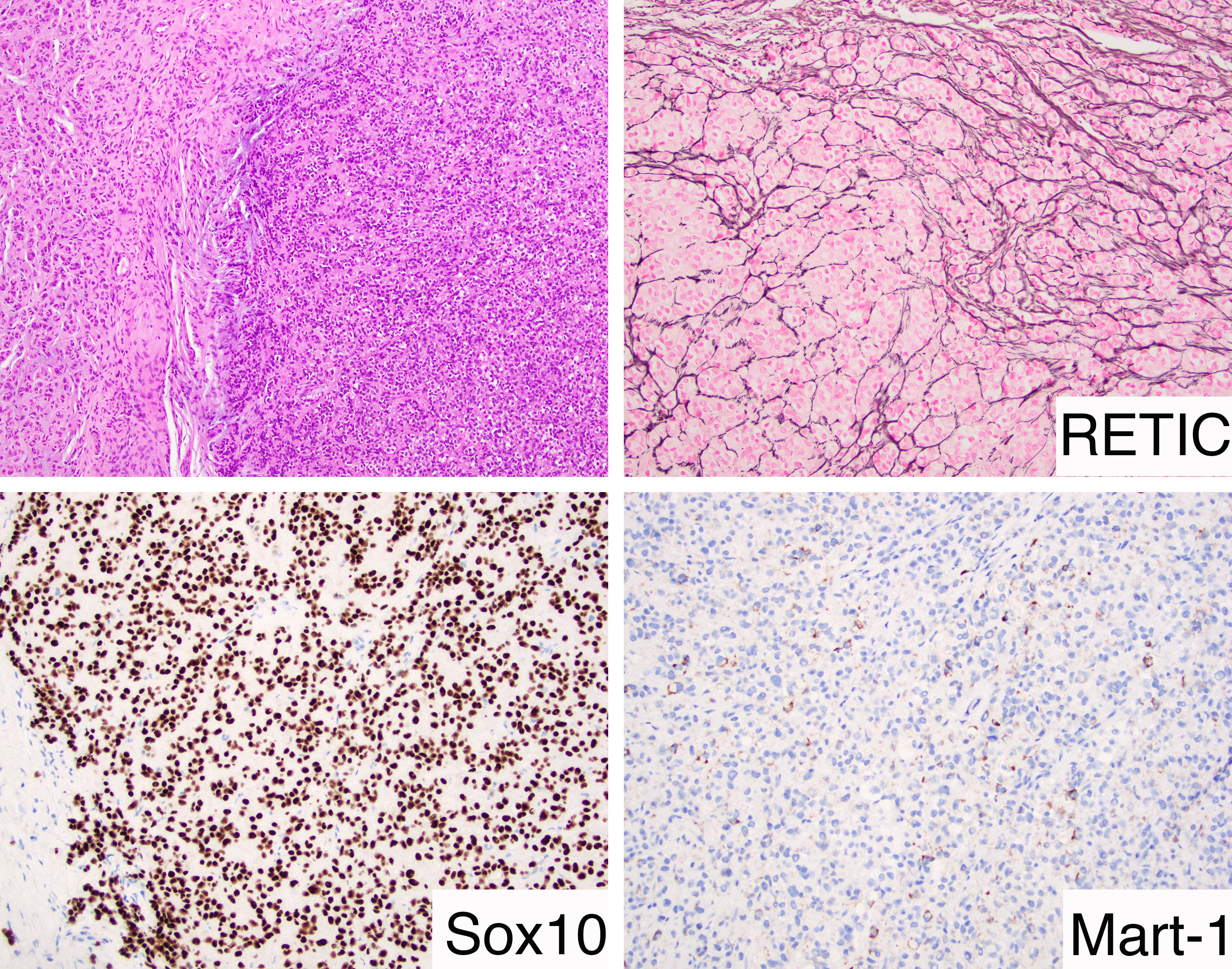
This challenging lesion was diagnosed with the help of Dr. Justin Bishop from UT Southwestern. The tumor shows a lobulated growth pattern and is composed of nests of epithelioid cells with minimal atypia and a rare mitotic figure. The nuclei show clumped chromatin and numerous pseudonuclear inclusions. Some cells show clear cell change. The complete immunohistochemical panel revealed that this tumor was negative for p63, 34BE12, Cam 5.2, HMB45, MiTF, GFAP and SMA. An Ini-1 immunostain showed preserved expression. Epithelioid Schwannomas (ES) are rare benign soft tissue tumors and even in cases with atypical features lesions do not metastasize and have a low recurrence rate1.
Option A: The lack of overt atypia and mitotic activity in addition to the presence of a basal membrane around nests (highlighted by the humble Reticulin histochemical stain) as well as the lack of diffuse staining for HMB45 help to rule out melanoma. Interestingly, focal weak Mart-1/Melan A has been reported to be positive in 1 case from a series of 24 ES lesions, a potential pitfall1.
Option B: Negativity for several myoepithelial markers (p63, Keratins, GFAP, SMA) helps rule out a myoepithelial lesion.
Option D: Finally, ECMT, a rare lesion that exhibits the RREB1-MKL2 fusion, tends to never occur outside the anterior tongue, shows nodules of spindle-stellate cells in a reticular net-like pattern and show prominent myxoid stroma and is usually GFAP and Keratin positive2.
References
1 - Hart J et al. Epithelioid Schwannomas: An Analysis of 58 Cases Including Atypical Variants. Am J Surg Pathol. 2016 May;40(5):704-13.
2 - Dickson BC et al. Ectomesenchymal Chondromyxoid Tumor: A Neoplasm Characterized by Recurrent RREB1-MKL2 Fusions. Am J Surg Pathol. 2018 Oct;42(10):1297-1305.
Case contributed by Carlos Prieto-Granada, M.D., Assistant Professor, Anatomic Pathology, UAB Department of Pathology