Case History
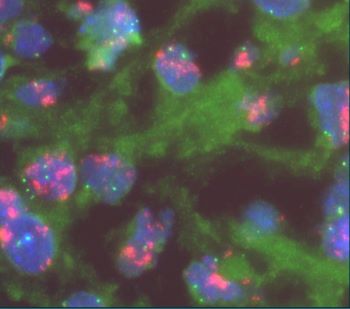
55 YOM presented with thigh mass. A biopsy was performed and showed the following lesion illustrated in the images. FISH analysis using a dual-color MDM2/CEN12 probe [Red=MDM2; Green=CEN12] was performed.
What is the diagnosis?
- Myxoid liposarcoma
- Myxofibrosarcoma
- Low-grade fibromyxoid sarcoma
- Dedifferentiated liposarcoma


Answer D: Dedifferentiated liposarcoma
Dedifferentiated liposarcoma may arise de novo or in association with atypical lipomatous tumor/well-differentiated liposarcoma. The risk of dedifferentiation varies with location of primary tumor. Subcutaneous neoplasms rarely undergo dedifferentiation whereas the highest risk is observed in tumors of intra-abdominal and retroperitoneal locations. An increase in tumor growth is typically seen in cases that arise in the setting of well-differentiated liposarcoma.
Histologically, dedifferentiated areas can be of high or low grade including area that resembles undifferentiated pleomorphic sarcoma, with pleomorphic spindle, polygonal, and multinucleated cells or with marked inflammation, or an area resembling inflammatory myofibroblastic tumor. Occasionally, the dedifferentiated component may resemble myxoid soft tissue neoplasm exhibiting myxoid stroma and delicate fibrovascular network entering the differential diagnosis with myxoid liposarcoma, myxofibrosarcoma, and low-grade fibromyxoid sarcoma. If present, the well-differentiated (lipogenic) component in dedifferentiated liposarcoma is mostly discrete showing an abrupt transition from the dedifferentiation component, but the two components can be intermingled in a mosaic pattern. Nonetheless, in small biopsies, only one component may predominate.
Similar to atypical lipomatous tumor/well-differentiated liposarcoma, supernumerary ring and giant marker chromosomes are the hallmarks of dedifferentiated liposarcoma detected in up to 90% of the cases. Ancillary study with immunohistochemical is positive for CDK4 and MDM2 (12q13-15 region) in nuclei of tumor cells in both the well-differentiated and the dedifferentiated component. Amplification of these genes can be detected by molecular genetic techniques. FISH analysis with dual-color probes is typically used to detect copy number changes in MDM2 gene and used to compare to CEN12 gene. A ratio of MDM2:CEN12 of more than 2 is interpreted as amplified MDM2. In addition to copying number changes in 12q13-15, chromosomal microarray shows recurrent gains in 12q24, 20q13, 6q22-q24, 9q33-q34, and losses in 13q14-q21, 11q22-q23.
Dedifferentiated liposarcoma is treated with complete surgical resection with negative margins whereas surgical debulking is typically used for cases of large multifocal retroperitoneal/intra-abdominal tumors. Anatomic location is considered the most important prognostic factor, in which, retroperitoneal/intra-abdominal tumors exhibit the worst clinical outcome with the highest rate of local recurrence. Up to 20% of dedifferentiated liposarcoma show distant metastases with an overall mortality of 25-30% at 5-year follow-up.
Selected references:
1. Dedifferentiated liposarcoma. WHO classification (5th edition). 2019
2. Kobayashi A, Sakuma T, Fujimoto M, Jimbo N, Hirose T. Diagnostic Utility and Limitations of Immunohistochemistry of p16, CDK4, and MDM2 and Automated Dual-color In Situ Hybridization of MDM2 for the Diagnosis of Challenging Cases of Dedifferentiated Liposarcoma. Appl Immunohistochem Mol Morphol. 2019;27(10):758-763. doi:10.1097/PAI.0000000000000677
3. Thway K, Jones RL, Noujaim J, Zaidi S, Miah AB, Fisher C. Dedifferentiated Liposarcoma: Updates on Morphology, Genetics, and Therapeutic Strategies. Adv Anat Pathol. 2016;23(1):30-40. doi:10.1097/PAP.0000000000000101