Case History
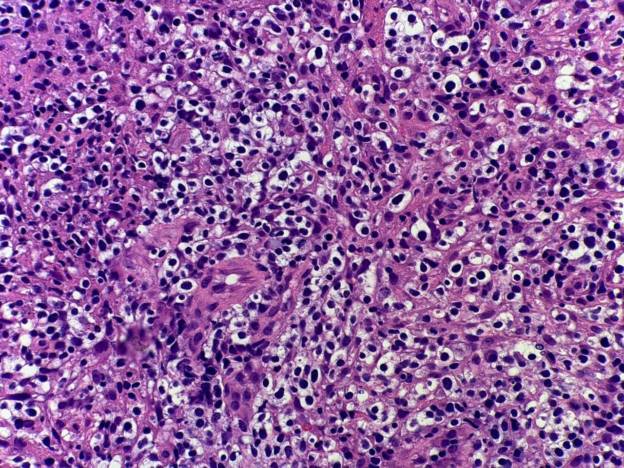
A 53-year-old female with acute hypoxic respiratory failure and pancytopenia. Imaging showed pulmonary infiltrates and lymphadenopathy. The morphology of a right axillary lymph node and immunohistochemical stains are shown.
What is the diagnosis?
A. Follicular lymphoma
B. Angioimmunoblastic T-cell lymphoma
C. Anaplastic large T-cell lymphoma (ALCL)
D. Burkitt lymphoma
Correct answer: B. Angioimmunoblastic T-cell lymphoma
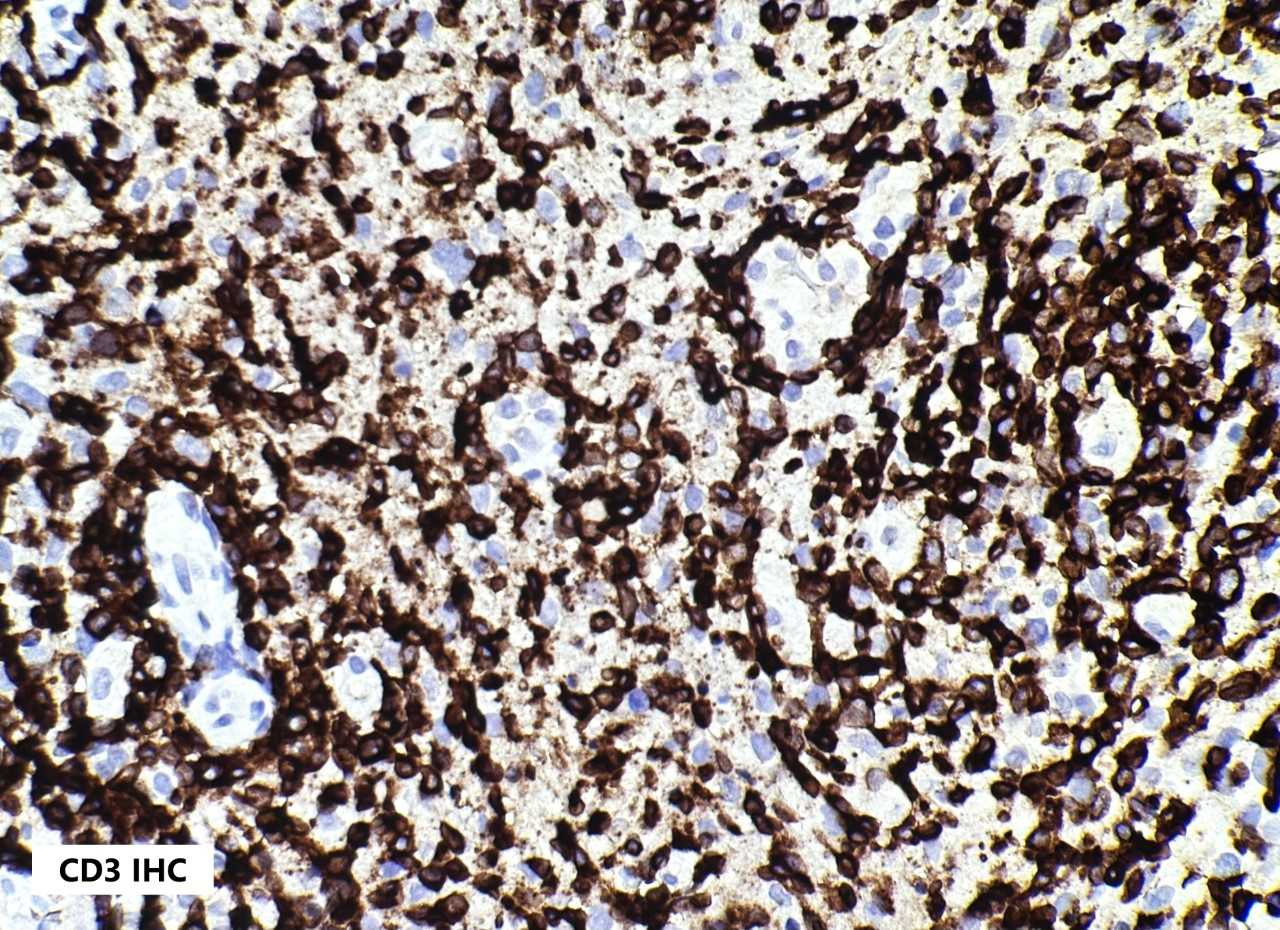
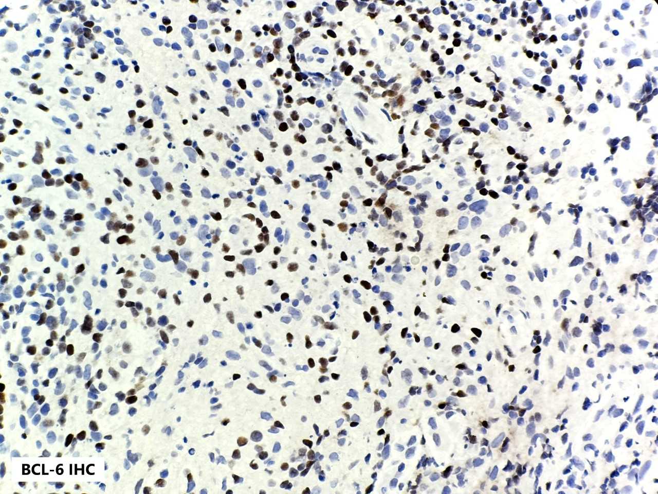
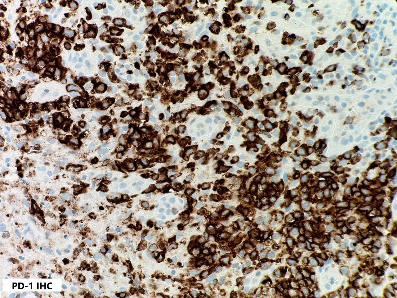
The H&E section shows diffuse involvement by heterogenous atypical lymphocytes with moderate amounts of clear cytoplasm and prominent vascular proliferation. These cells are positive for CD4, CD2, CD3, CD5 and T-follicular helper markers including CD10, BCL6, ICOS and PD-1. Angioimmunoblastic T-cell lymphoma is a member of the T-follicular helper family of nodal T-cell lymphomas and is characterized by a T-cell infiltrate with expression of at least two T-follicular helper markers, high endothelial venule proliferation, and follicular dendritic network expansion.
Case contributed by: Sarah DePew, D.O., Hematopathology Fellow, UAB Pathology