Case History
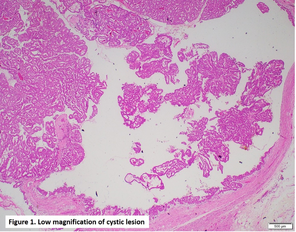
A 66-year-old female presented due to a cystic lesion of the pancreatic head approximately 5.6 cm. A Whipple procedure was performed and 5.7 cm-sized ill-defined multiloculated cystic mass involving the main pancreatic duct was identified by gross examination. Figures include H&Es, immunohistochemical stain (HepPar-1), and albumin mRNA in situ hybridization (ISH).
What is the diagnosis?
A. Pancreatic ductal adenocarcinoma
B. Metastatic hepatocellular carcinoma
C. Intraductal oncocytic papillary neoplasm
D. Intraductal tubulopapillary neoplasm
Correct Answer: C. Intraductal oncocytic papillary neoplasm
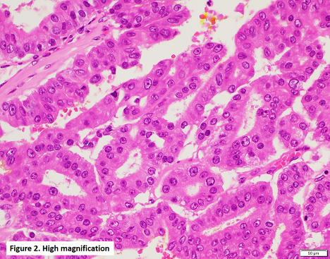
Intraductal oncocytic papillary neoplasm (IOPN) of the pancreas is a subtype of IPMN and histologically characterized by papillary projections (Figure 1) lined by oncocytic cells with abundant granular eosinophilic cytoplasm and large, fairy uniform nuclei containing single, prominent nucleoli (Figure 2). Of note, IOPNs may automatically be graded as high-grade dysplasia.
IOPNs are genetically distinct from classic, mucin-producing IPMNs since they lack KRAS and GNAS mutations commonly found in IPMNs. Interestingly, IOPNs show DNAJB1-PRKACA fusion, previously reported in cases with fibrolamellar hepatocellular carcinoma.
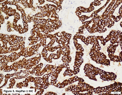
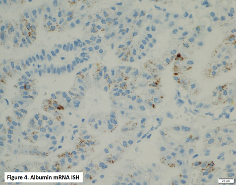
IOPNs express HepPar-1 (Figure 3) by IHC and albumin mRNA by ISH (Figure 4), which may help distinguish them from other subtypes of IPMN.
Concerning patients’ long-term outcomes, IOPNs demonstrate indolent behavior even in the presence of invasive carcinoma, which occurs in about 25-61% of IOPNs.
References:
- Vyas M et al. DNAJB1-PRKACA fusions occur in oncocytic pancreatic and biliary neoplasms and are not specific for fibrolamellar hepatocellular carcinoma. Mod Pathol. 2020 Apr;33(4):648-656.
- Wang T et al. Intraductal Oncocytic Papillary Neoplasms: Clinical-Pathologic Characterization of 24 Cases, With An Emphasis on Associated Invasive Carcinomas. Am J Surg Pathol. 2019 May;43(5):656-661.
Case contributed by: Goo Lee, M.D., Ph.D., Associate Professor, Anatomic Pathology, UAB Pathology