Case History
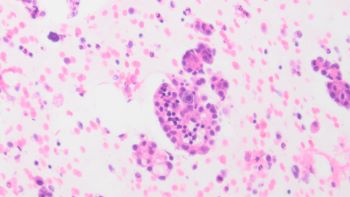
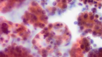
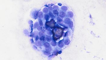
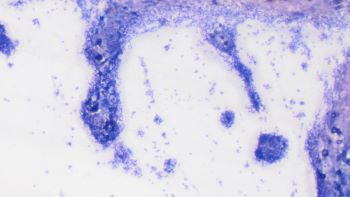
The patient is a 73-year-old female with history of complex cystic and solid ovarian mass on CT. A pelvic wash was performed, exhibiting multiple clusters as seen in the pictures below (Pap stain, Diff-Quik and cell block).


What IHC panel would be most appropriate to clarify the lesion's nature?
A. MOC31, BerEP4, PAX8, Claretinin
B. Calretinin, WT1, BAP1
C. SOX10, MART1, S100
D. CD56, Chromogranin, synaptophysin
Correct answer: A. MOC31, BerEP4, PAX8, Claretinin
Discussion:
When it comes to body fluids, one of the most common, yet challenging, differentials is between carcinoma or mesothelioma. The pelvic wash in question shows multiple clusters of epithelioid cells, with enlarged nuclei and prominent nucleoli both on Pap stain and DIff Quik, as does in the cell block. Other features observed were mitotic figures and areas of calcification. The clinical presentation of ovarian mass, makes the diagnosis of mesothelioma less likely, favoring the diagnosis of carcinoma cells present in the pelvic wash. Therefore, the most adequate panel would be MOC31, BerEp4, Pax8 and calretinin. BerEp4 and MOC31 are both anti-EpCAM antibodies, highlighting the membrane stain, as targets the epithelial cell adhesion molecule, positive in carcinomas. PAX 8 is a member of paired box gene family of transcription factors involved in the development of multiple organs, including organs from the Mullerian ducts, with a nuclear staining pattern. Calretinin is a calcium binding protein positive in both normal and neoplastic mesothelial cells. Its presence in the panel is justified, as a negative result will reinforce the epithelial nature of the malignant cells. WT1, even though is a marker for serous carcinoma of the ovary, it is also positive in malignant mesothelioma, and therefore a positive result would not contribute to solve tthe differential. Even though melanoma is an universal mimicker, the clinical presentation and the absence of classic cytomorphologic features justify not including the markers on panel C on an initial approach. The cells have no neuroendocrine features to justify the panel on option D.
Case contributed by: Igor Damasceno Vidal, M.D., Cytopathology Fellow, UAB Pathology