Case History
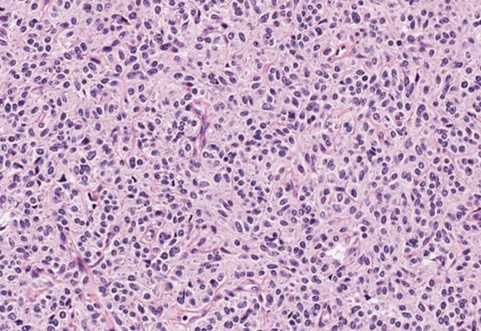
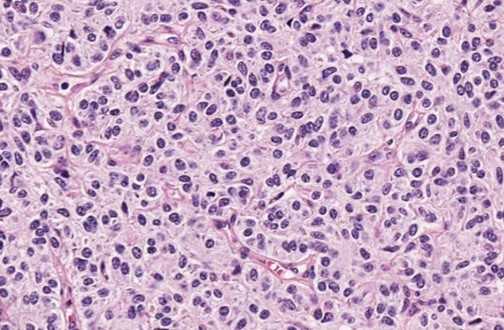
A 63-year-old female presented with a history of coughing, wheezing, and recurrent pneumonia and underwent CT imaging for evaluation of her symptoms. A 1.4 cm central, well-circumscribed nodule with increased attenuation was identified on contrast CT.


What is the diagnosis?
A. Lymphoma
B. Carcinoid tumor
C. Pulmonary paraganglioma
D. Solitary fibrous tumor
Correct answer: B. Carcinoid tumor
Discussion:
Carcinoid tumors (CTs) are neuroendocrine malignancies in the lung. Depending on the location, patients can experience a variety of symptoms. Central CTs may present with coughing, wheezing, and recurrent pneumonia due to the bronchial obstruction. Peripheral CTs are generally asymptomatic and found incidentally on imaging. There are no risk factors recognized for CTs. There are two subtypes: typical and atypical. These tumors have a neuroendocrine morphology with organoid patterns of growth with many types of arrangements (trabecular, rosette, insular, ribbon, follicular, etc). Tumor cells are uniform with finely granular nuclear chromatin, moderate to abundant eosinophilic cytoplasm, inconspicuous nucleoli, and granular chromatin. Differences between the two subtypes are that CTs have < 2 mitoses/2 mm2 and lack necrosis, while atypical CTs have 2–10 mitoses/2 mm2 and/or foci of necrosis (usually punctate). CTs are positive for LMW cytokeratins and neuroendocrine markers. TTF1 tends to be positive in peripheral tumors but negative in central tumors.
Case contributed by: Sarah Anderson, D.O., Forensic Pathology Fellow, UAB Pathology