 |
Gorgas Case 2021-03 |
 |
|
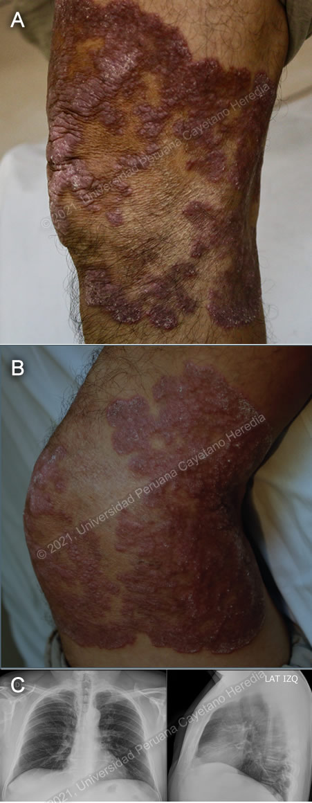
The following patient was seen as an outpatient at the dermatology clinic at Hospital Cayetano Heredia. We would like to thank Dr. Ramos and Dr. Bravo for their assistance with this case.  History: A 55-year-old man presents with a history of illness of approximately 40 years, which started with a non-painful, non-pruritic, small elevated erythematous lesion over his left knee which slowly grew over the decades. He treated it with topical antibiotics and corticosteroids on several occasions, with no improvement. He denies any other symptoms including neuropathy. He has had no other skin problems in the past 40 years that he can recall. Epidemiology: Born in Chiclayo, a city on the northern coast of Peru. He moved to Lima approximately 30 years ago and works as an electrician. Denies any recent travel. Denies past medical or surgical history, does not recall if he received BCG vaccine. Denies contact with TB patients. No notable family history of infections or immunodeficiencies. Physical Examination: Examination of the skin revealed an erythematous, serpiginous, verrucous plaque in the extensor surface of the knee which extended to the sides, with central atrophy, some hyperalgesia and preserved sensation (Images A and B). Joint mobility was unaffected. No lymphadenopathy. The rest of the exam was unremarkable. Imaging studies: CXR was normal (Image C). Laboratory Examination: Hb: 14.7 g/dL; Hct. 44%; WBC 4 550 (neutrophils: 62%, eosinophils: 1, lymphocytes: 30%); Platelets: 200 000. Gluc: 98 mg/dL, Urea: 17 mg/dL, Creat: 1.0 mg/dL, AST 22 U/L, ALT 17 U/L, GGT 31 U/L, albumin 4.2 g/dL.
|
|
Diagnosis: Cutaneous tuberculosis due to Mycobacterium tuberculosis - Lupus vulgaris form 
 Discussion: Tuberculin skin testing (TST) was positive (15mm), and a skin biopsy revealed tuberculoid granulomas, with giant multinucleated cells and central caseation (Image D). These findings and the clinical presentation of the patient are sufficient to make a diagnosis of lupus vulgaris. Acid-fast stain of the sputum was negative for mycobacteria. Cultures are pending. Cutaneous tuberculosis may also be classified depending on the number of mycobacteria that can be identified from the lesions. Paucibacillary forms include tuberculosis verrucosa cutis and lupus vulgaris. Multibacillary forms include primary inoculation tuberculosis, scrofuloderma, tuberculosis cutis orificialis, acute miliary tuberculosis, and tuberculous gumma. Diagnosis is a challenge, as lupus vulgaris is paucibacillary and demonstration of acid-fast bacilli is often not possible. Histopathology of a skin biopsy reveals tuberculoid granulomas with or without caseation, with few or no bacilli. Mycobacterial cultures are usually negative, however, TSTs are usually positive. PCR for mycobacteria is accurate and rapid, and even allows for differentiation of MTB from other non-tuberculous mycobacteria if the right primers are chosen, but requires high-complexity laboratories and skilled technicians (https://onlinelibrary.wiley.com/doi/abs/10.1046/j.1365-4362.2003.01461.x). |