UAB is opening a new, state-of-the-art zebrafish research facility.
 |
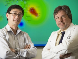
| Sam Cartner, assistant vice president for Animal Research Services, and Mary-Ann Bjornsti, chair of Pharmacology & Toxicology, were among many faculty and staff members instrumental in the creation of UAB’s new zebrafish lab. |
“The zebrafish shares 90 percent of the genome with humans and has become the No. 3 model for the NIH behind mice and rats,” says Stephen Watts, Ph.D., professor of biology and co-director of the facility. “With this new facility, the elements are here to form a research group.”
The co-principal investigators of the HSF-GSF grant include Sam Cartner, Ph.D., assistant vice president for Animal Research Services and director of the Animal Resources Program; Mary-Ann Bjornsti, Ph.D., chair of Pharmacology & Toxicology; and Susan Farmer, D.V.M., Ph.D., senior clinical veterinarian in Animal Resources, and Watts.
Leaders from the schools of Medicine and Dentistry, UAB Comprehensive Cancer Center and the College of Arts & Sciences comprise an oversight committee that will ensure all investigators have access and that infrastructure needs are met, Bjornsti says.
Cartner says equipment and technical support will be available to all UAB investigators using the facility.
“The researchers this will benefit have immediate needs to further their research in a variety of diseases,” Cartner says. “A zebrafish facility within the UAB biomedical research community is a key step.”
The facility initially will provide critical infrastructure needed for research programs in developmental biology, genetics, metabolism and cancer. This also will enhance the efforts of current UAB scientists who are poised to apply this model to high-priority studies of tumor promotion, skeletal abnormalities and nutrition.
“We have had interest from 10 investigators,” Cartner says, “and we expect more to come forward.”
Why zebrafish?
The zebrafish was determined to be a model system for studying vertebrate development and genetics almost 40 years ago.
Since that time, zebrafish embryos have become very popular worldwide as a means of understanding how all vertebrates — including people — develop from the moment that sperm fertilizes an egg.
The eggs are clear and develop outside the mother’s body, enabling scientists to watch a zebrafish egg grow into a newly formed fish under a microscope. Researchers can watch in real time while the cells divide and form different parts of the fish’s body.
In the span of two to four days, zebrafish can form eyes, heart, liver, stomach, skin and ultimately grow into a newly formed fish. Watts has watched all of this from a small satellite facility he has been maintaining for several campus researchers.
“Their regeneration time is very short,” Watts says. “I can get 100 eggs every other day from a female zebra fish, and I can watch full development of every major organ and every major component necessary under a microscope.”
Because zebrafish are closely related to humans, they are more likely to be similar to us in many biological traits than a more distantly related organism. These traits include genes, developmental processes, anatomy, physiology and behaviors.
There are other advantages:
- Transparent embryos enable researchers to monitor the behavior of single cells in live embryos and watch the cells divide and trace each cell and its offspring in the making of the complete organism.
- Single cells or groups of cells can be transplanted into host embryos, which allows investigators to analyze the behavior of cells at different stages or ask how mutant cells behave in wildtype embryos.
- These are less expensive models to house and maintain long term.
Recruitment tool
Thirteen entities have committed significant funds to the project: Office of the Vice President for Research and Economic Development; the schools of Medicine and Dentistry; College of Arts & Sciences; Comprehensive Cancer Center; Alabama Drug Discovery Alliance; Center for Clinical and Translational Sciences; Nutrition and Obesity Research Center; Comprehensive Arthritis Center; Cystic Fibrosis Research Center; and the departments of Pharmacology & Toxicology, Cell Biology and Genetics.
The facility also is considered a recruitment tool. Two researchers — John Parant, Ph.D., assistant professor of Pharmacology & Toxicology, and Peter Jezewski, D.D.S., Ph.D., assistant professor of Dentistry — were successfully recruited to UAB this past fall in part because of the new facility.
“The need to build a facility to recruit top faculty was a driving force,” says Bjornsti.
Watts says he and his colleagues have extensive expertise in aquatic animal husbandry. They will be responsible for optimizing use along with facility co-director Susan Farmer, D.V.M., Ph.D., senior clinical veterinarian in Animal Resources.


